
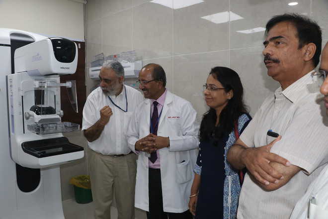
PGI gets state-of-the-art 3D mammography machine

Tribune News Service
Chandigarh, August 31
Prof Jagat Ram, Director, PGI, inaugurated the latest generation digital breast tomosynthesis (DBT) mammography machine. The machine is also called 3D mammography and has been installed in radiology section of the New OPD. There are very few such machines available so far, stated Prof MS Sandhu, Head, Department of Radiodiagnosis and Imaging.
The incidence of breast cancer is increasing rapidly in India. Mammography is the earliest and the best imaging modality for screening and diagnosis of breast cancer. The sensitivity of conventional mammography for the detection of breast cancer varies with breast density and is lower for women with dense breasts. These women need additional investigations like ultrasound as a dense breast can either obscure a lesion or can misinterpret a lesion when no lesion is present.
The DBT image helps in these patients by minimising the tissue overlap that can hide cancers. In addition, DBT uses low dose X-ray system and computer reconstruction to create 3D images. In breast tomosynthesis, X-ray tube moves in an arc over the compressed breast capturing multiple images of each breast from different angles so that the entire breast is better evaluated. These digital images are then reconstructed into a set of 3D images by a computer for correct interpretation, hence improving the accuracy of the diagnosis and reducing the false positive rates. Thus, the need for breast biopsy, which is an invasive procedure, also reduces in certain cases.
Breast tomosynthesis will greatly help in early detection and better diagnosis of patients with breast-related symptoms as well as in screening mammography. It will also reduce the need for additional ultrasound required for the mammography in certain patients. The system also has a computer-aided lesion detection system, which further helps in the diagnosis, especially for residents-in-training.
“This mammography machine has a stereotactic biopsy system with which we can do image-guided biopsy for breast lesions and for breast microcalcification, which will also greatly benefit patient care in the tricity and in the surrounding areas,” said Prof Veenu Singla and Dr Tulika Singh.
Prof Sandhu said the machine comes with a specimen radiography unit, which is the first to be installed in India. This would greatly help in conducting advanced research on breast cancer patients for evaluation of positive tumour margins, which would benefit patient care. It also provides additional information in case of early breast cancer in biopsy specimen for better visualisation of calcification, thus reducing the chances of repeating the biopsy and increasing the confidence level. The Director also planted some saplings outside the mammography unit as part of the nationwide tree plantation drive for Green India.
Unlock Exclusive Insights with The Tribune Premium
Take your experience further with Premium access.
Thought-provoking Opinions, Expert Analysis, In-depth Insights and other Member Only Benefits
Already a Member? Sign In Now