Berlin, August 23
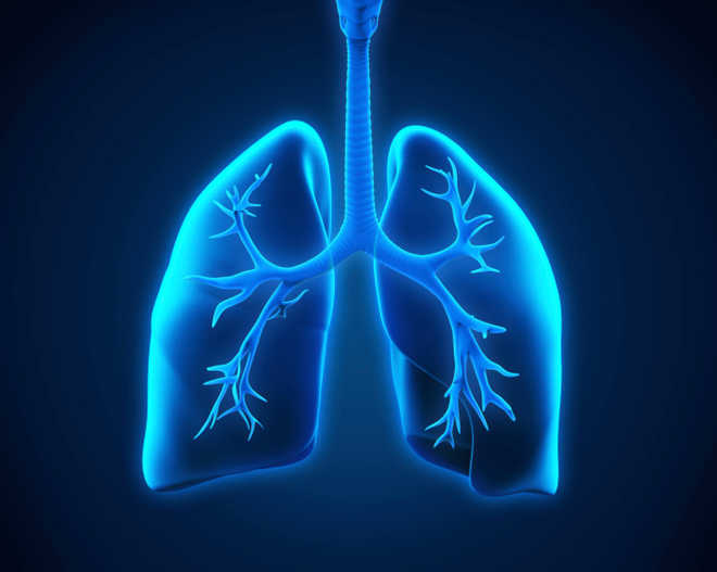
Using a novel technique which enables high resolution imaging of damaged lung tissues, scientists have found the changes caused by severe COVID-19 in the structure of the organ's blood vessels and air sacs, findings that may support the development of new treatment methods against the disease.
In the study, published in the journal eLife, the scientists developed a new X-ray technique which enables high resolution and three-dimensional imaging of lung tissues infected with the novel coronavirus SARS-CoV-2.
Using the new method, the researchers, including those from the University of Gottingen in Germany, observed significant changes in the blood vessels, inflammation, and a deposition of proteins and dead cells on the walls of the lungs' tiny air sacs called alveoli.
They said these changes make gas exchange by the organ either difficult or impossible.
According to the scientists, the new imaging approach allows these changes to be visualised for the first time in larger tissue volumes, without cutting and staining, or damaging the tissue.
They said it is particularly well suited for tracing small blood vessels and their branches in three dimensions, localising cells of the immune systems present at inflammation sites, and measuring the thickness of the alveolar walls.
Due to the three-dimensional reconstruction of the lung tissues, the researchers said the data could also be used to simulate gas exchange in the organ.
Since X-rays penetrate deep into tissue, they said scientists can use the method to understand the relation between the microscopic tissue structure and the larger function of an organ.
"Based on this first proof-of-concept study, we propose multi-scale phase contrast X-ray tomography as a tool to unravel the pathophysiology of COVID-19," the researchers wrote in the study.
The scientists believe the technique will support the development of treatment methods, and medicines to prevent or alleviate severe lung damage in COVID-19, or to promote recovery.
"It is only when we can clearly see and understand what is really going on, that we can develop targeted interventions and drugs," said Danny Jonigk, a co-author of the study from Medical University Hannover in Germany. PTI
Join Whatsapp Channel of The Tribune for latest updates.


